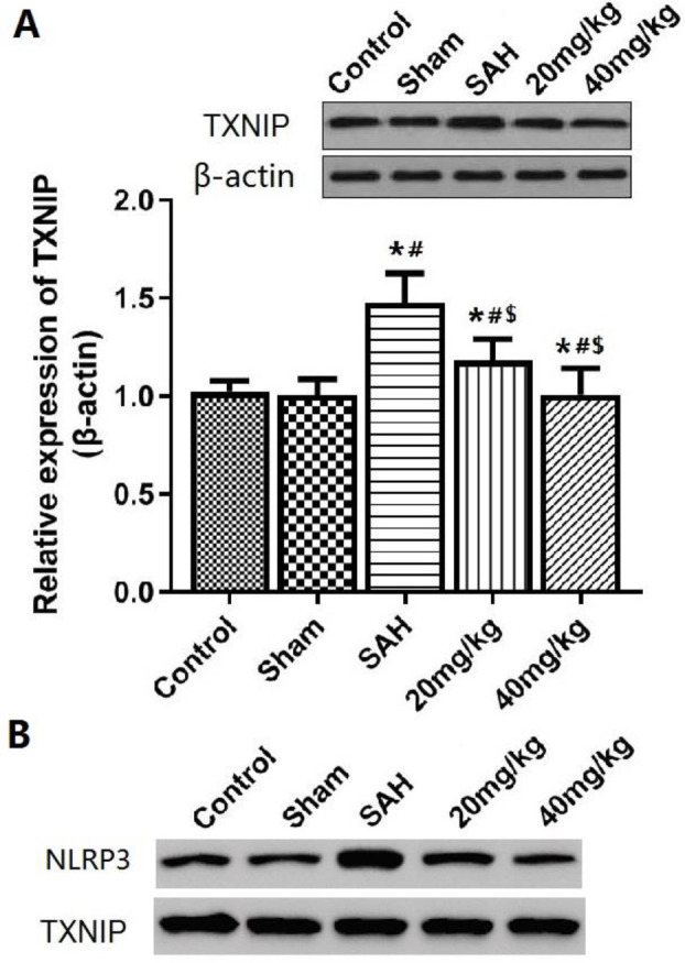
Figure 6.
TXNIP expression and TXNIP-NLRP3 interaction in brain tissue after SAH

A: Expression of TXNIP in brain tissue analyzed by Western Blot; B: Immunoprecipitation analysis of the interaction of TXNIP and NLRP3 in brain tissue. *P<0.05 vs control group, #P<0.05 vs Sham group, $P<0.05 vs SAH group
TXNIP: thioredoxin interacting protein; NLRP3: NOD like receptors pyrin domain-containing 3; SAH: subarachnoid hemorrhage
