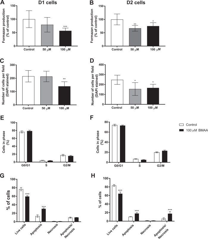
Fig. 2. Effects of BMAA on neural stem cell viability and proliferation in daughter cells after one (D1) or two passages (D2).
Cell viability was determined using the MTT assay (A, B) and proliferation by counting the number of cells using DAPI staining (C, D) in daughter cells of neural stem cells treated with 50 or 100 µM BMAA. The cell cycle phase was analyzed by flow cytometry (E, F), apoptotic and necrotic cells were assessed with the annexin V-PI assay (G, H) in daughter cells of neural stem cells exposed to 100 µM BMAA. Values represent mean ± SD from three independent experiments, each with six replicates. Statistically significant differences from control are indicated as follows: *p < 0.05, **p < 0.01 and ***p < 0.001 (one-way ANOVA followed by Tukey–Kramer test, or Student’s t-test when comparing only two groups).

