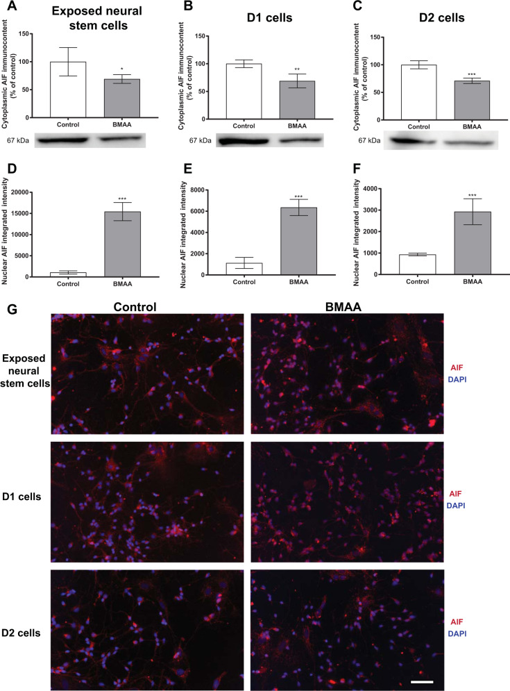
Fig. 3. Involvement of the apoptosis-inducing factor (AIF) in cell death triggered by BMAA in neural stem cells.
AIF levels were evaluated by western blot in cytoplasmic homogenates from neural stem cells exposed to 250 µM BMAA (A), or D1 (B) and D2 (C) daughter cells of neural stem cells exposed to 100 µM BMAA. β-tubulin was used as a loading control. Representative blots of three experiments are shown. Nuclear AIF levels were analyzed by immunocytochemistry using ImageJ (D–F). Representative images are shown (G). Values represent mean ± SD from three independent experiments. Statistically significant differences from control are indicated as follows: *p < 0.05, **p < 0.01 and ***p < 0.001 (Student’s t-test). Scale bar: 30 µm.

