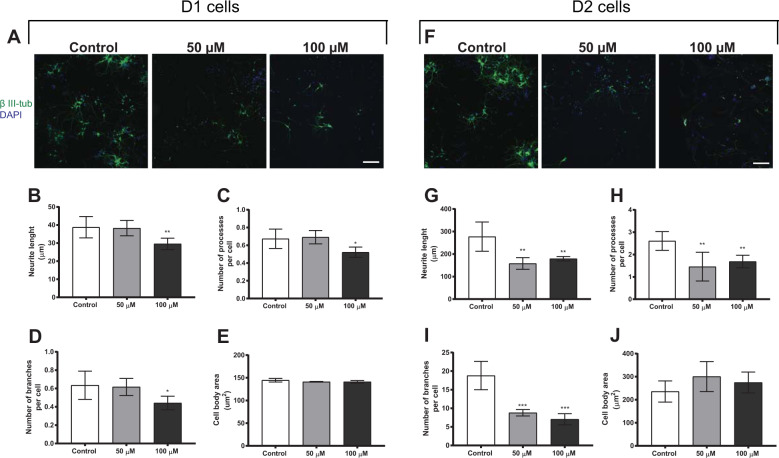
Fig. 6. Morphometric alterations in neurons derived from daughter cells (D1 and D2) of neural stem cells treated with 50 or 100 µM BMAA.
Representative images of cells immunostained with anti-β III-tubulin (green) and DAPI (blue) (D1 cells, A; D2 cells, F). Morphometric analysis was conducted using an ImageXpress Micro XLS Widefield HCA System (Molecular Devices, Sunnyvale, CA, USA), where images were automatically captured and analyzed with the MetaXpress Software. Neurite length (B, G), the number of processes per cell (C, H), the number of branches per cell (D, I), and the cell body area (E, J) were determined. Values represent mean ± SD from three independent experiments, each with six replicates. Statistically significant differences from control are indicated as follows: *p < 0.05, **p < 0.01 and ***p < 0.001 (one-way ANOVA followed by Tukey–Kramer test). Scale bar = 50 µm.

