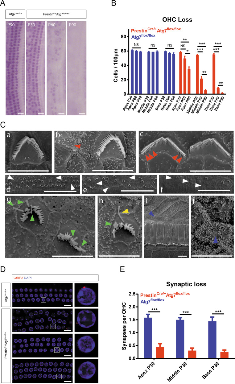
Fig. 3. Morphological changes in PrestinCre/+Atg7flox/flox mice and controls.
A Representative hematoxylin staining images of the cochlear middle turn depicting the gradual loss of OHCs over time. B The average number of OHCs per 100 μm dropped by P30 and dramatically decreased at P60 and P90, especially in the basal turns. C SEM images from control mice (a) and Atg7-knockout mice (h–j). (b) A bundleless OHC (red arrowhead). (c) Stereocilia were missing from the inner row (red arrowheads), while the regular V/W-shaped staircase structure was maintained. (d–f) Three consecutive fields of an apical turn, within which part c was closer to the middle turn and part a was more apical. OHCs of all three rows randomly lost their hair bundles (white arrowheads). (g, h) At P60, the stereocilia were degenerated. Splayed stereocilia and persistent kinocilia were identified (green and yellow arrowheads, respectively). (i, j) The dead OHCs were replaced by supporting cells with microvilli (blue arrowheads). D, E Immunolabeling of CtBP2 (red channel) showed the loss of presynaptic ribbons of OHCs. Insets to the right represent typical OHCs with or without ribbons. Z-stack quantification of CtBP2 puncta per OHC showed significant differences between Atg7 knockouts and controls. N = 6 for each group. Scale bar: A: 20 μm; C(a–c, g–j): 5 μm; C(d–f): 50 μm; D: 10 μm.

