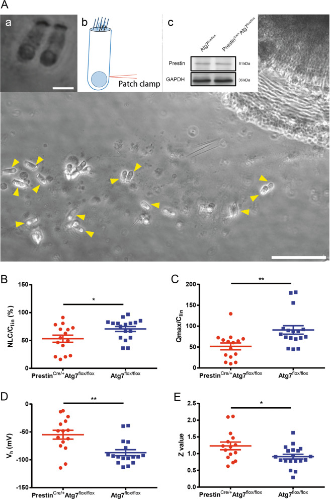
Fig. 5. OHC electromotility was disturbed in PrestinCre/+Atg7flox/flox mice.
A OHCs were dissected from the whole-mount cochleae, and HCs were indicated by yellow arrowheads. OHCs were differentiated from IHCs based on their characteristic morphology and unique electromotility, and two of them are shown in a. b shows the position where the electrode was patched onto the OHC body. Western blot showed the prestin expression in PrestinCre/+Atg7flox/flox mice at P30 (c). B–E NLCs recorded were pooled and normalized to the corresponding Clin. Qmax, Clin, Vh, and z were obtained from a curve fit of the NLC response for each OHC, as shown by scatter plots of individual data and normalized mean values with standard errors. Significant differences for these parameters were seen between the two groups. Atg7-mutant OHCs: N = 15; the control OHCs: N = 17 (western blot: N = 3 for each group). Scale bar: A: 100 μm; a: 10 μm.

