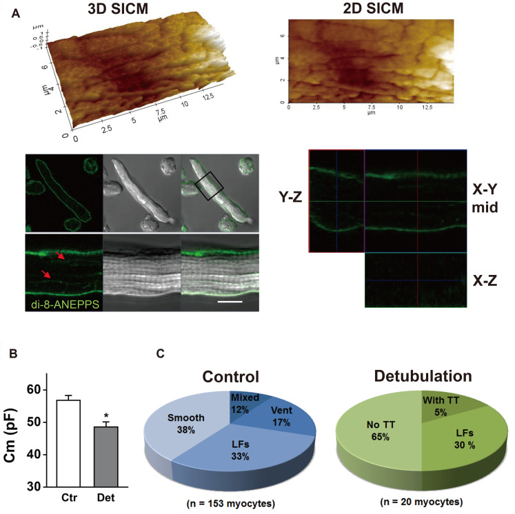
Fig. 7. Validation of formamide-induced detubulation in the atrial myocytes.
(A) Atrial cells were stained with di-8-ANEPPS after formamide-induced detubulation procedure. Confocal and surface scanning ion conductance microscopy (SICM) images were taken from the same scanning areas of the cell. SICM images of the detubulated cell surface show the existence of longitudinal fissures (LFs). A plane of the di-8-ANEPPS confocal image is overlaid with an optical transmission image, with an indication box of the scanning area. Arrowheads in the confocal image indicate presumable LF membranes. Representative X–Y, X–Z, and Y–Z confocal plane images are presented. (B) Detubulation reduced the cell membrane capacitance (*p < 0.05). (C) Proportions of each subtype, classified by SICM surface structure, are compared between control and detubulation cells.

