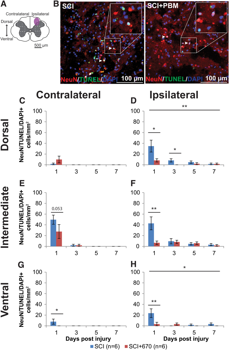
FIG. 5.
Neuronal cell death occurs early following spinal cord injury, which is reduced by 670 nm treatment. (A) The schematic representation of the spinal cord illustrates the dorsal, intermediate, and ventral regions of interest for analysis (enclosed by dashed lines, 0.1 mm2). Approximate location of injury epicenter is indicated by the purple shaded area. (B) Example images of NeuN (red), TUNEL (green), and DAPI (blue) triple-positive cells from SCI untreated and light-treated groups ipsilateral to the injury at dorsal region at 1 dpi. (C,D) Quantification of NeuN+TUNEL+DAPI+ cells, expressed as triple-positive cell density within the region of interest, in the dorsal region of the spinal cord, contralateral (C) and ipsilateral (D) to the injury of untreated and light-treated groups. (E,F) NeuN+TUNEL+DAPI+ cell density in the intermediate regions of interest contralateral (E) and ipsilateral (F) to the injury. (G,H) NeuN+TUNEL+DAPI+ cell density in the ventral regions of interest contralateral (G) and ipsilateral (H) to the injury. Data are expressed as mean ± SEM; n values indicated (legend) are for each time-point. Statistical comparisons between SCI and SCI+670 (LMER) are shown; *p < 0.05, **p < 0.01. dpi, days post-injury; LMER, linear mixed-effects models; SCI, spinal-cord injured animals without red-light treatment; SCI+670, spinal-cord injured animals with red-light treatment; SEM, standard error of the mean. Color image is available online.

