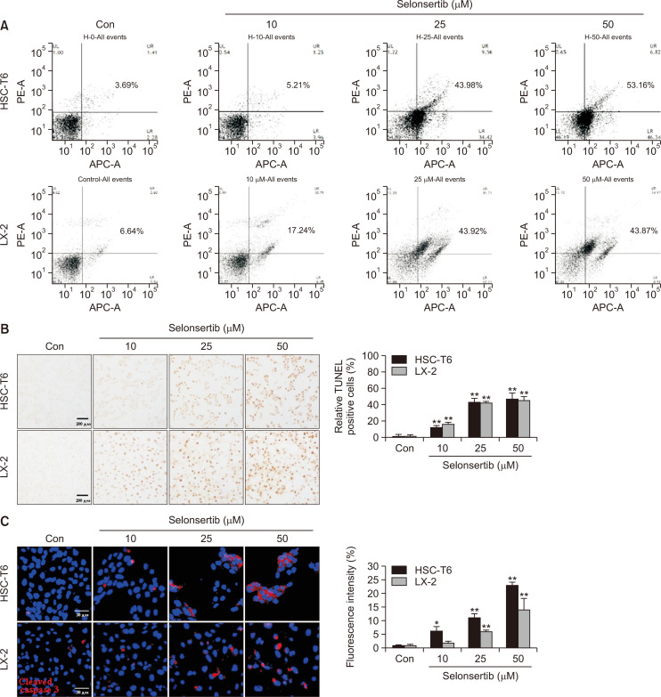
Fig. 2.
Effect of selonsertib on HSC apoptosis. (A) HSC-T6 and LX-2 cells were treated with various concentrations of selonsertib (10-50 μM) for 48 or 72 h. The HSCs were double-stained with annexin V-APC and propidium iodide (PI) and analyzed by the FACS-verse flow cytometer. Annexin V-APC events represent apoptotic cells for early stage (low right quadrants) and late-stage (upper right quadrants). The analysis was conducted with two independent experiments. (B) The induction of apoptosis by selonsertib (10-50 μM) was observed by TUNEL staining (200× magnification). Data are represented by fold changes of TUNEL-positive cells. (C) Red fluorescence intensity from cleaved caspase-3 staining after the treatment of selonsertib. Data are shown as the fold changes compared to the control cells. All the values are presented as mean ± SD. *p<0.05 and **p<0.01 versus the control.

