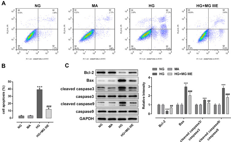
Figure 3.
MG IIIE attenuated the cell apoptosis of HG-induced podocytes. (A) Apoptotic cells were detected via flow cytometry. (B) The apoptotic rate of podocytes was quantified. (C) The expression of apoptosis-related proteins were evaluated using Western blot analysis. ***P<0.001 vs. MA; ##P<0.01, ###P<0.001 vs. HG.
Abbreviations: MG IIIE, Mogroside IIIE; NG, normal glucose; HG, high glucose; MA, mannitol.

