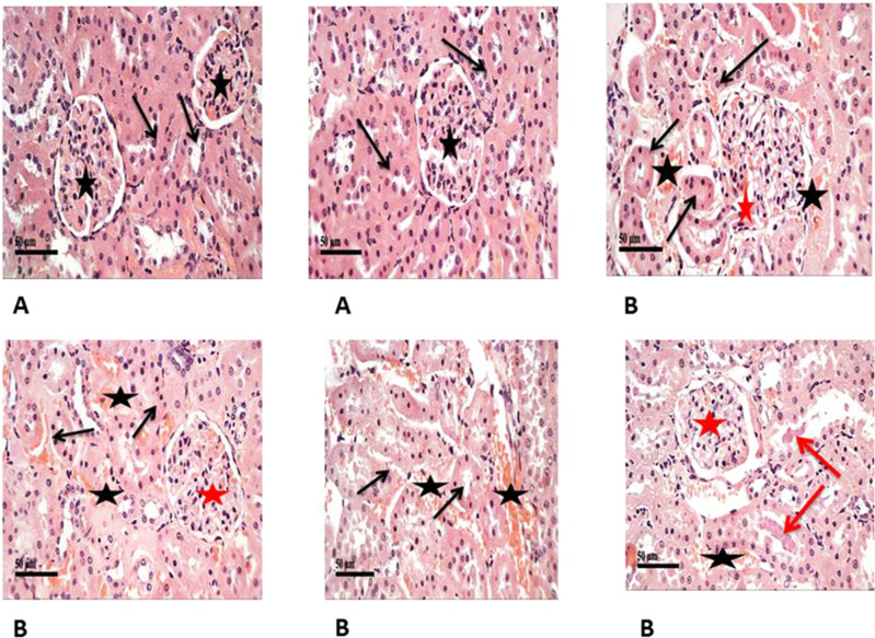
Figure 3.
Shows H&E staining of histopathological changes in the groups of kidney tissues (400×). (A) Control samples had normal morphological features of renal parenchyma with apparent intact renal corpuscles (stars) and different nephron segments displaying apparent intact renal tubular epithelium (arrows) as well as intact vasculature. (B) Model samples showed the effect of STZ on histological structure of kidneys showed marked congestion of renal glomeruli (red stars) as well as many dilated and congested intertubular blood vessels (black stars). Frequent records of degenerative or necrotic changes of renal tubular epithelium in proximal and distal convoluted tubules with significant records of pyknotic nuclei (black arrows) and occasional intraluminal eosinophilic casts (red arrows).

