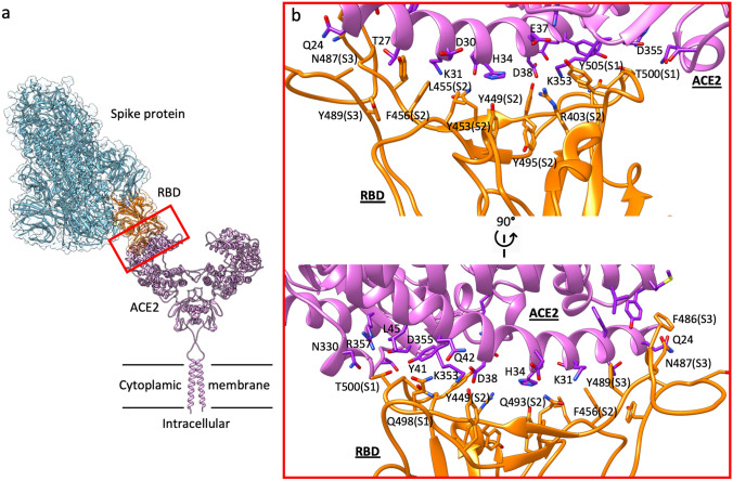
Fig. 1.
Supramolecular organization of the spike protein:ACE2 complex and the RBD:ACE2 binding surface. a Schematic representation showing the dimeric ACE2 protruding from the cytoplasmic membrane and acting as receptor for the trimeric SARS-CoV2 spike protein in prefusion open conformation, which binds through one of the three available RBD in up position. b two side-views of the contact interface between the RBD (orange) and ACE2 (purple). The three RBD sites (S1, S2, and S3) primarily responsible for binding ACE2 α1 helix are indicated within parentheses

