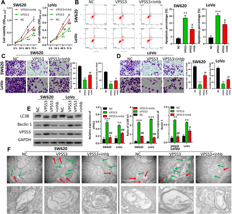
Figure 3.
VPS53 inhibited CRC cell carcinogenesis by regulating autophagy. SW620 and LoVo cells were treated with the VPS53-overexpressed plasmid and the autophagy inhibitor (Inhb). (A) CCK-8 assay indicates the influence of Inhb on the VPS53-inhibited proliferation of SW620 and LoVo cells, **P < 0.01 vs NC group; #P < 0.05, ##P < 0.01 vs VPS53 group. (B) The role of Inhb on the VPS53-induced apoptosis analyzed by flow cytometry, and the apoptosis percentage is shown, **P < 0.01, ***P < 0.001 vs NC group; #P < 0.05 vs VPS53 group. (C and D) VPS53 plasmid and Inhb-treated SW620 and LoVo cells were subjected to the migration and invasion assays, and the migrated and invaded cells were counted, **P < 0.01, ***P < 0.001 vs NC group; #P < 0.05, ##P < 0.01 vs VPS53 group. (E) Western blot analysis results show expressions of LC3B and Beclin 1, and the quantitative results were analyzed, **P < 0.01, ***P < 0.001 vs NC group; #P < 0.05, ##P < 0.01 vs VPS53 group. (F) Ultrastructure of autophagic bodies observed by an electron microscope. The red arrow denotes autophagosomes and the green arrow denotes the lysosomes.

