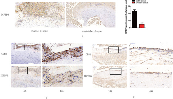
Fig. 8.

Immunochemistry analysis of IGFBP6 in histologically stable and unstable plaques
A. IGFBP6-positive area (brown) is significantly decreased in the unstable plaque compared with that in the stable plaque (×10) (****p < .0001) B. Colocalization of CD68+ macrophages in the border between the fibrous cap and necrotic core, in the area where IGFBP6 is expressed (×10). The black rectangle indicates the local enlarged view (×40). C. CD31+ endothelial cells are colocalized with IGFBP6 (× 10). The black rectangle indicates the local enlarged view (×40).
