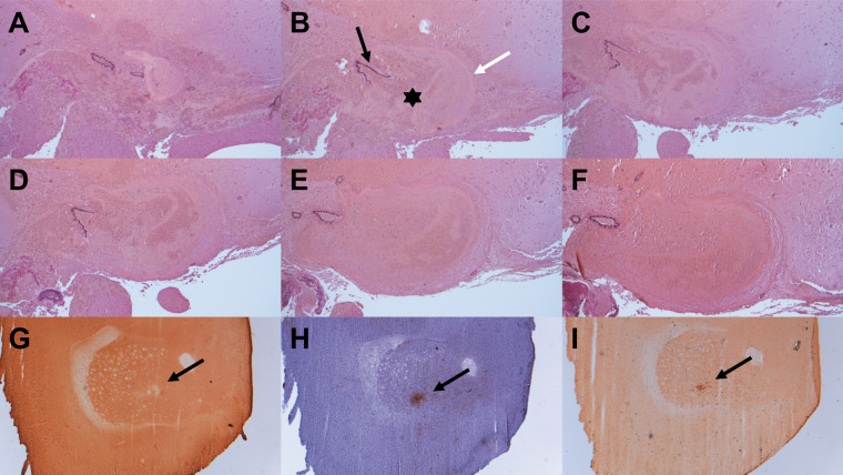
Figure 6.
Histological analysis confirmed pseudoaneurysm observed in DSA (a–f, elastica van gieson, 100× magnification): a round structure, filled with an appreciable amount of erythrocytes (b – asterisk), surrounded by a fibrinogen coat (b – white arrow) and in some sections in contact with a sustentative vessel (b – black arrow). Initially, the nearby vessel is closed (a), afterwards the ruptured vessel section is visible (b–e) and at the last slice, the vessel is intact again (f). Additionally, histology showed smaller infarctions of the striatum after staining with MAP2 (g; black arrow, 25× magnification). Microglia and macrophages were found in the CD68-staining (i; black arrow, 25× magnification) and microglia in Iba1-staining (h; black arrow, 25× magnification).

