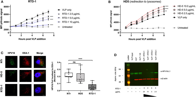
Figure 2.
RTD-1 treatment of HPV16 results in significantly reduced uptake and localization of virions to early endosomes highlighted by sparse detection of large aggregates at cell surfaces and reduced cell binding capacity of RTD-1 treated virions. Time course of pHrodo-HPV16 VLP internalization and trafficking to low pH endosomal compartments in the presence of (A) RTD-1 or (B) HD5. Shown is the mean pHrodo signal intensity of triplicate wells ± SD. Background signal from untreated cells included. (C) Immunofluorescence imaging of internalized HPV16 PsV untreated (NTr) or incubated with 5.0 μg/ml RTD-1 or HD5. (Left) Representative images from each field of view per treatment group (scale bar = 10 μm). (Right) Quantitation of HPV16 and EEA-1 colocalization was performed using ImageJ with the JaCOP plugin using 10 individual fields of view (10–20 cells/field) from two independent experiments. (D) Western blot of cell surface bound HPV16 L1 capsid protein in the presence of RTD-1. Membrane fractions are shown, probed for HPV16 L1 (green) and actin (red). ns = not significant, **p < 0.01, ****p < 0.0001 two-way ANOVA followed by Dunnett's multiple comparison test against untreated VLPs at each timepoint for uptake experiments. One-way ANOVA followed by Dunnett's multiple comparison test against untreated PsV in colocalization studies.

