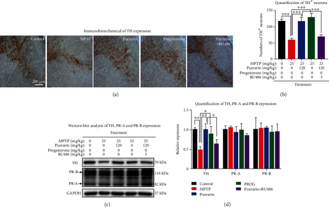
Figure 2.

Puerarin protected dopaminergic neurons from MPTP-induced damage in mice. (a) Immunohistochemical staining of TH expression. The frozen brain sections were analyzed by immunohistochemical staining with anti-TH antibody. The images were captured under an Olympus microscope (Tokyo, Japan). (b) Quantification for TH+ neurons. The number of TH+ neurons in each group (n = 3) were counted. ∗∗∗p < 0.001. (c) Western blot analysis of TH, PR-A, and PR-B expression. Midbrain tissues were collected and analyzed by Western blotting with antibodies against TH, PR-A, and PR-B, whereas GAPDH was analyzed as control. Representative blots for each group were shown. (d) Quantification of TH, PR-A, and PR-B expression. Western blots (n = 3) in (c) were determined by a densitometric method. ∗p < 0.05, ∗∗p < 0.01.
