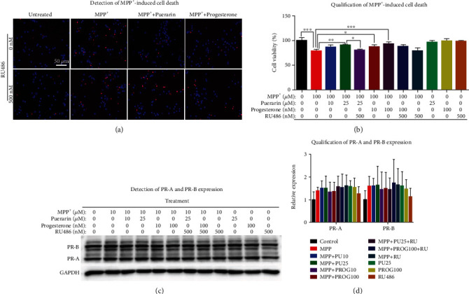
Figure 4.

Puerarin enhanced the survival of primary midbrain neurons against MPP+-induced neurotoxicity. (a) Detection of MPP+-induced cell death. Primary midbrain neurons were treated with MPP+, puerarin, progeresterone, and RU486 as indicated. Cell viability was assessed by Hoechst 33342 and PI staining. The representative images were shown. (b) Quantification of neuronal viability. Hoechst- and PI-positive cells were counted under a Zeiss fluorescence microscope (Carl Zeiss, Jena, Germany). ∗p < 0.05; ∗∗p < 0.01; ∗∗∗p < 0.001. (c) Western blot analysis of PR-A and PR-B expression. Primary midbrain neurons were treated with MPP+, puerarin, progeresterone, and RU486 as indicated. The expression of PRs was analyzed by Western blot analysis with specific antibodies, while GAPDH was analyzed as the loading control. Representative blot was shown. (d) Quantification of PR-A and PR-B expression. Western blots (n = 3) in (c) were determined by a densitometric method. ∗p < 0.05.
