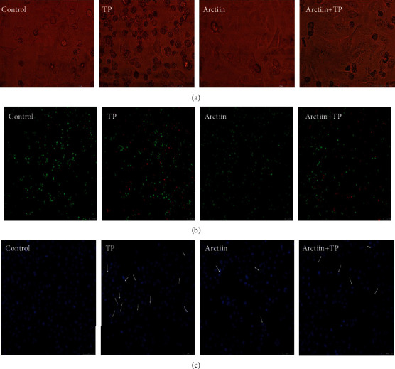
Figure 2.

Effects of TP, arctiin, and TP + arctiin on the morphology of HepG2 cells. Treated cells were visualized using (a) light microscope and fluorescence microscope after stained with (b) Calcein-AM/PI mixed dye and (c) Hoechst 33258. White arrows indicated the condensation and fragmentation of chromatin.
