Figure 6.
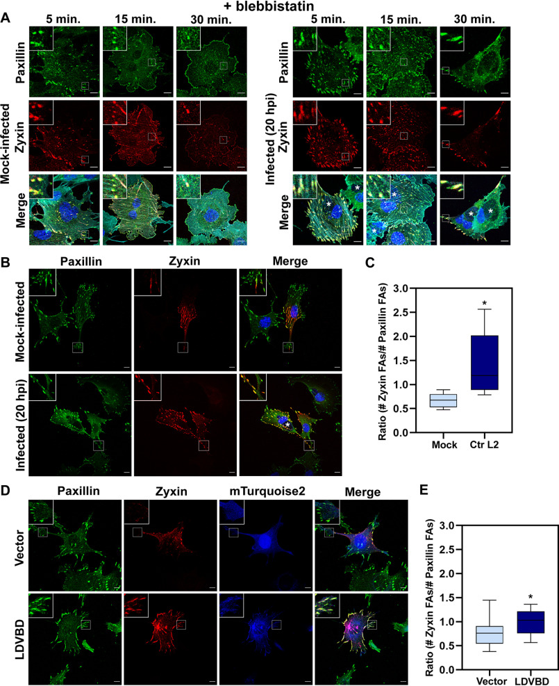
An increased number of FAs contain the maturation marker zyxin in Chlamydia-infected and LDVBD-expressing cells. A, MEF cells were transfected with RFP-zyxin via electroporation and then mock-infected or infected with CtrL2 for 20 h. Cells were treated with 10 μm blebbistatin and fixed at the indicated time points and then stained for the focal adhesion marker paxillin (green) as well as actin (cyan; shown in composite images). Chlamydia-containing inclusions were visualized by staining with DAPI and are marked with asterisks in composite images. B, CtrL2-infected MEF cells expressing RFP-zyxin were processed for immunofluorescence with anti-paxillin antibody to visualize focal adhesions. Inclusions were visualized by staining with DAPI. Representative images are shown. Scale bar, 10 μm. C, the paxillin and zyxin channels were submitted to the Focal Adhesion Analysis Server to obtain a mask of each channel. The focal adhesion number was then counted using the particle-counting plug-in from ImageJ. Data are the number of zyxin adhesions present divided by the number of paxillin adhesions present per individual cell and illustrated as a box-whisker plot. Whiskers represent the lowest and highest data point still within 1.5 times the interquartile range. For statistical analyses, ANOVA was used to determine significance when compared with a mock control (*, p < 0.001) (n = 10 cells). D, MEF cells expressing RFP-zyxin as well as vector only or LDVBD-mTurquoise2 were processed for immunofluorescence with anti-paxillin antibody. Representative images are shown. E, quantification performed as described in C. For statistical analyses, ANOVA was used to determine significance when compared with vector-only control (*, p < 0.05) (n = 10 cells).

