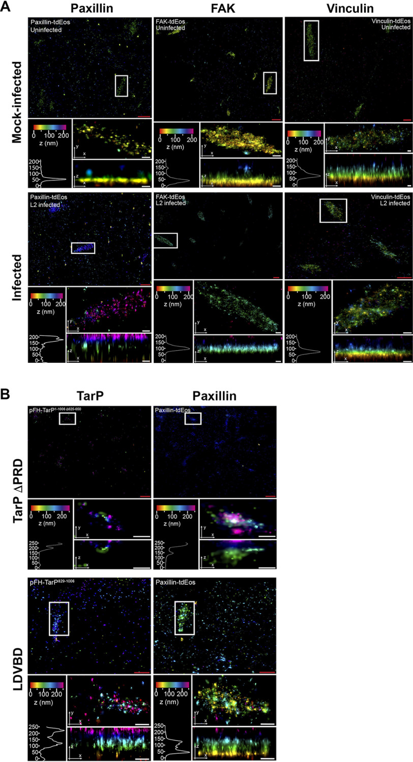
Figure 7.
TarP-targeted focal adhesions display altered nanoscale architecture. A, COS7 cells were pretransfected with paxillin-tdEos, FAK-TdEos, or vinculin-TdEos on gold fiducial coverslips and were mock-infected or C. trachomatis–infected for 20 h. The cells were fixed and processed for iPALM imaging. Representative images are shown from n = 3. For each sample, multiple panels are provided. The top panel shows the top view of the area around the focal adhesion of interest (white border). The middle panel displays a top view of the focal adhesion indicated by the white border. The bottom panel shows the side view and corresponding z histograms. Note the significant shifts in paxillin and FAK localization, but not vinculin. B, COS7 cells were co-transfected with paxillin-tdEos and either TarP ΔPRD or LDVBD only by electroporation. The cells were seeded on gold fiducial coverslips and processed for iPALM at 24 h post-transfection at n = 2. Description of each panel is as above in A. Note the significant shift in the location of paxillin within the TarP-positive focal adhesions. The various colors indicate the distance (z-coordinates) from the gold fiducial marker (e.g. z = 0 nm; red). Red scale bar, 1 μm. White scale bar, 200 nm.

