Figure 1.

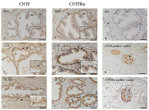
Immunohistochemistry localization of CNTF and CNTFRa in prostate samples. CNTF is highly expressed in basal layer (arrows) of BPH (a), CYP (b) and NL-RP (c) while the secretory layer (arrowheads) is mainly negative. The stromal tissues are weakly stained for CNTF in all samples analysed. CNTFRa is highly expressed in basal layer (arrows) of BPH (d), CYP (e) and NL-RP (f) while the other tissues are mainly negative. The basal layer of glandular epithelium is identified by p63 marker (arrow, g). Pictures in h) and i) show a ganglion (arrow) positive for CNTF (h) and for CNTFR (i) used as positive internal controls. The insets show higher magnification of the area indicated by asterisk, scale bars: 30 μm. Scale bars: a,b,c,d,f,g) 100 μm; e,h,i) 200 μm.
