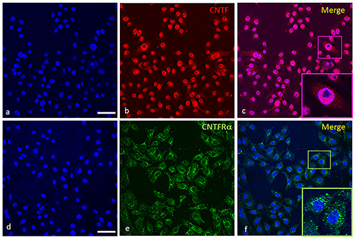
Figure 2.

Immunofluorescence of CNTF and CNTFRɑ in PWR-1E cell line. In (a) and (d) are stained the nuclei in blue. CNTF (b, red staining) is localized mainly in nuclei while the cytoplasm is weakly stained for CNTF (b,c). In (c), the nucleoli are negative as depicted in the inset (c, Merge). CNTFRɑ (e, green staining) is localized in the cytoplasm of the cells as shown in (f ) and it is especially intense in the perinuclear region (see inset in f, Merge). Scale bars: a,b,c,d,e,f ) 50 μm; Insets in c,f ) 17 μm.
