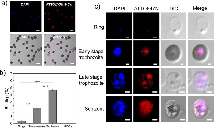
Figure 2.
Analysis of the binding of Glc-NCs to different blood stages of P. falciparum. (a) CLSM imaging of ATTO@Glc-NCs incubated for 15 min with infected RBCs showed specific binding of intracellular P. falciparum (red channel, ATTO647N; blue channel, DAPI; scale bar: 5 μm). (b) FACS analysis of the binding of ATTO@Glc-NCs incubated for 15 min with highly synchronized P. falciparum cultures. Parasitemia was determined using SYBR Green (FITC channel), and ATTO647N dye was detected to determine the binding of ATTO@Glc-NCs (APC channel). ****P ≤ 0.0001. (c) CLSM imaging of individual asexual erythrocyte stages incubated with ATTO@Glc-NCs showed specific binding for all the stages (red channel, ATTO647N (ATTO@Glc-NCs); blue channel, DAPI; scale bar: 2 μm).

