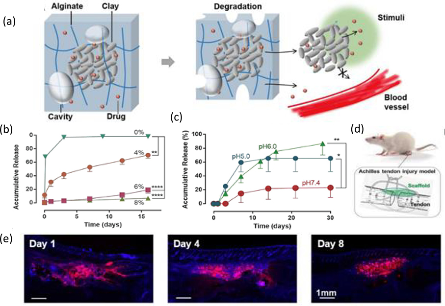
Figure 6. Composite hydrogel with high loading and local release of IGF1.
(a) Clay composite microparticles containing Laponite nanoparticles (represented by gray discs) and adsorbed drug molecules (red spheres) localized within a biodegradable alginate network containing micro-sized basins (gray ovals) and free drug (not sequestered within the microparticle). (b) In vitro drug release profiles from composite hydrogels containing various percentages of clay in cPBS medium at a pH of 7.4. (c) In vitro drug release profiles from 6% clay composite hydrogels within release medium at different values of pH. (d) Use of drug-releasing scaffolds (green) to treat Achilles tendon injury in a rat model. (e) DAPI staining (blue) of tissue sections to examine the distribution of the fluorescein-labeled protein (red) as it changes over time in vivo. Adapted with permission.[112] Copyright 2018, WILEY-VCH.

