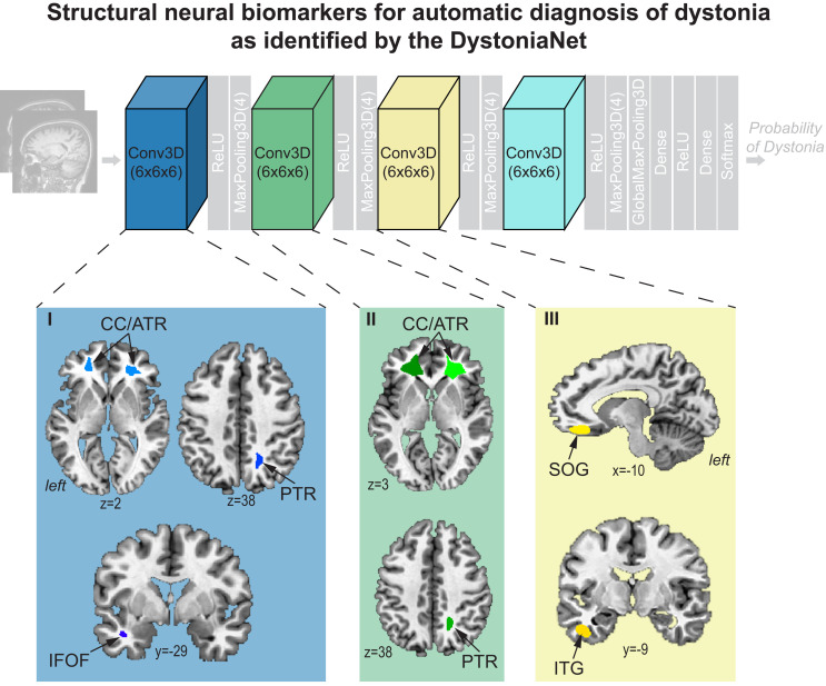
Fig. 2.
Microstructural neural network biomarker for automatic diagnosis of isolated dystonia as identified by the DystoniaNet platform. Brain regions as components of the biomarker are identified by the first three convolutional layers of DystoniaNet for diagnostic classification. Brain regions in the fourth layer are not visualized due to low spatial resolution. Axial and sagittal brain slices depict 2D visualizations of the most discriminative features in the AFNI standard Talairach–Tournoux space. ReLU, rectified linear unit; CC/ATR, corpus callosum/anterior thalamic radiation of corona radiata; PTR, posterior thalamic radiation of corona radiata; IFOF, inferior fronto-occipital fasciculus; SOG, superior orbital gyrus; ITG, inferior temporal gyrus.

