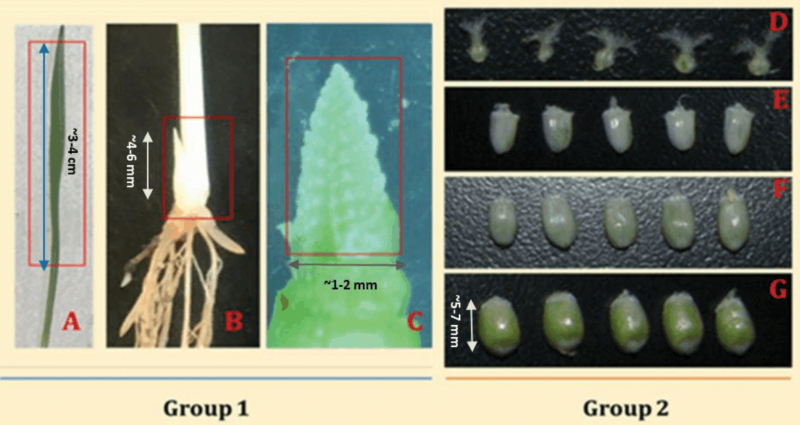
Figure 1.
Tissues sampled for the proteomics study, namely; (A) leaf, (B) tillering initiation site, (C) terminal spikelet stage, (D) Ovary, (E) 5 days post anthesis (5DAA), (F) 10 days post anthesis (10DAA), (G) 15 days post anthesis (15DAA). The seven target samples were categorized into Group 1, representing leaf, tiller initiation site, and the terminal spikelet sample while Group 2 represents ovary and progressive kernel development stages. For the leaf sample, the first leaf was sampled when the second leaf just emerging.

