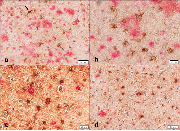
Figure 6.

Aggregatin expression in AD brains. (a) double immunolabeling of Aggregatin (brown) and Aβ (red). Arrows indicate reactive astrocytes with a binuclear morphology, (b) double immunolabeling of Aggregatin (brown) and Aβ (red) at higher magnification. The center of amyloid plaque is devoid of Aggregatin immunoreactivity, (c) double immunolabeling of Aggregatin (brown) and AT8 (red) at higher magnification. AT8-expressing intraneuronal inclusions are negative for Aggregatin immunoreactivity, and (d) double immunolabeling of Aggregatin (brown) and ApoE (red). Aggregatin-expressing reactive astrocytes are negative for ApoE immunoreactivity.
