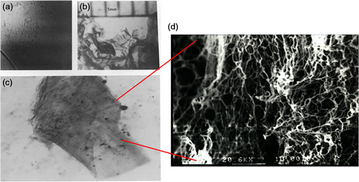
FIGURE 4.

Discovery of the first self‐assembling peptide scaffold material. (a) The structure was formed in phosphate‐buffered saline and transferred to a glass slide. The colorless membranous structures are isobuoyant; therefore, the image is not completely in focus (×75 Nomarski Phase contrast microscope.) (b) The structure stained bright red with Congo red and can then be seen by the naked eye (×15, each scale unit = 1 mm). (c) A portion of a well‐defined membranous structure with layers is clearly visible; the dimensions of this particular membrane are 2 × 3mm (×20)
