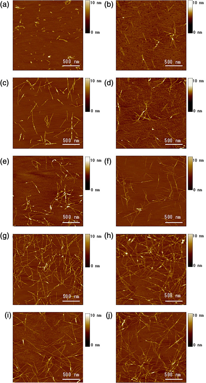
FIGURE 6.

Atomic force microscopy (AFM) images of RADA16‐I nanofiber at various time points after sonication. The observations were made using AFM immediately after sample preparation. (a) 1 min after sonication; (b) 2 min; (c) 4 min; (d) 8 min; (e) 16 min; (f) 32 min; (g) 64 min; (h) 2 hr; (i) 4 hr, and (j) 24 hr. Note the elongation and reassembly of the peptide nanofibers over time. By ~1–2 hr, these self‐assembling peptide nanofibers have nearly fully re‐assembled This process of sonication and re‐assembly can be repeated many times, thus truly demonstrating the natural power of self‐assembly. Image courtesy of Hidenori Yokoi 29
