
FIGURE 8.
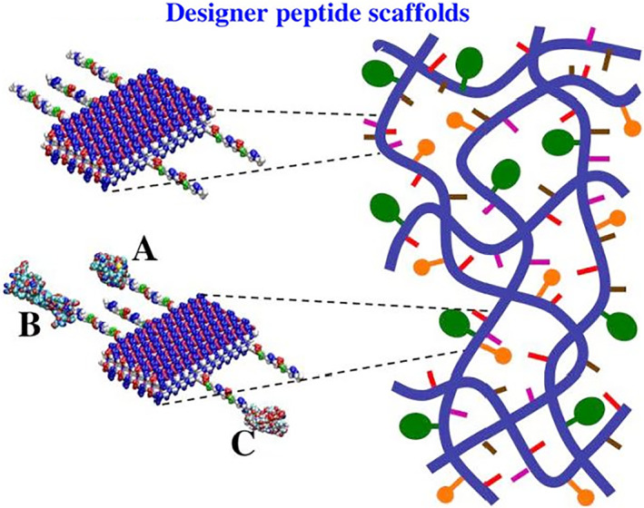
Molecular and schematic models of the designer peptides and of the scaffolds. Direct extension of the self‐assembling peptide sequence by adding different functional motifs. Blue thick lines represent the self‐assembling backbone and the yellow, pink, and tan lines represent various functional peptide motifs. Molecular model of a self‐assembling peptide nanofiber with functional motifs flagging from both sides of the double β‐sheet nanofibers. Either few or more functionalized and active peptide can be mixed at the same time. The density of these functionalized peptides can be easily adjusted by simply mixing them in various ratios, 1:1–1,000,000 or more, before assembling. They will then be part of the self‐assembled scaffold. The left panel shows enlargements of a small part of the peptide nanofibers on the right panel. The enlarged parts show more detail of co‐assembly of peptides with biologically active motifs, either different peptides (upper left panel), or different proteins (lower left panels: a, b, and c)