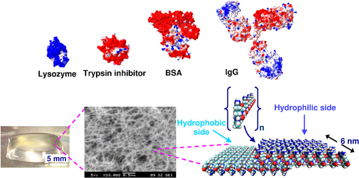
FIGURE 12.

Molecular representation of lysozyme, trypsin inhibitor, BSA, and IgG as well as of the Ac‐N‐(RADA)4‐CONH2 peptide monomer and of the self‐assembling peptide nanofiber. Color scheme for proteins and peptides: positively charged (blue), negatively charged (red) and hydrophobic (light blue). Protein models were based on known crystal structures from PDB (Protein Data Bank). Image courtesy of Sotirios Koutsopolous 43
