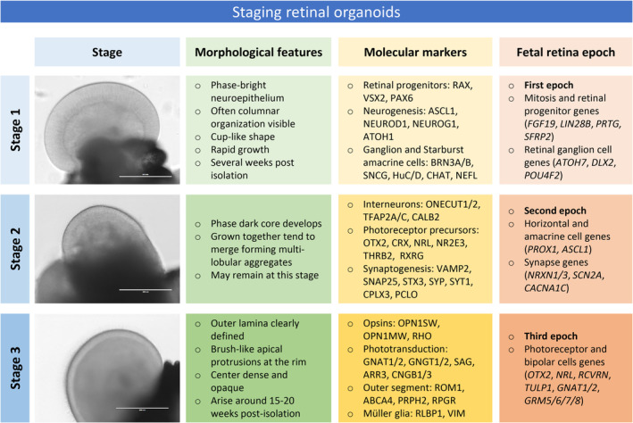
FIGURE 2.

Staging of retinal organoids. Based on histological features and molecular marker expression from organoid transcriptome studies, three major sequential stages of differentiation can be related to molecularly defined epochs in human fetal retina development. Bright‐field images on the left show retinal tissue in organoids at each stage (scale bar 400 μm). Table summarizes key morphological features, molecular markers, and corresponding human fetal development epoch at each organoid differentiation stage
