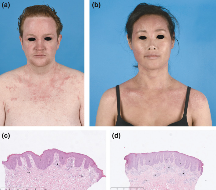
Figure 1.

(a, b) Clinical pictures of paradoxical erythema in a head and neck distribution with (c, d) corresponding histological haematoxylin and eosin staining of lesional biopsies. (a) Patient 1: a 28‐year‐old male patient showing a relatively sharply demarcated, minimally scaling, patchy erythema of the face and neck (scalp not affected), which was associated with burning and itching, but was notably different from his usual eczema. (b) Patient 2: a 29‐year‐old female patient showing a minimally scaling, patchy erythema of the face and neck (scalp not affected), which was asymptomatic. (c, d) Histological examination of lesional skin biopsies of patients 1 (c) and 2 (d), both obtained from the neck, revealed psoriasiform epidermal hyperplasia with bulbous elongated rete ridges (*), increased numbers of ectatic capillaries in the papillary dermis (→) and a moderate perivascular lymphocytic infiltrate (▲). Interestingly, spongiosis was largely absent in all biopsies.
