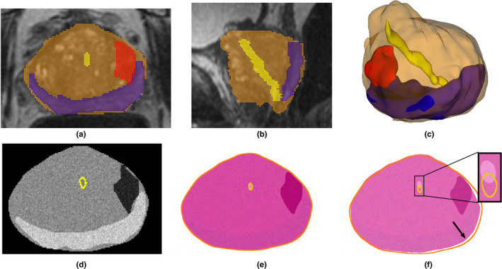
Fig. 2.

Radiology‐pathology digital phantom. The expert annotation of the prostate (orange), peripheral zone (blue), urethra (yellow), and cancer(red) on three‐dimensional (3D) T2 magnetic resonance imaging (MRI), shown in the (a) axial; (b) sagittal; (c) 3D views, were used to create a digital phantom of the prostate: (d) slice in the MRI phantom, (e) corresponding slice in the pathology phantom, and (f) imperfect corresponding slice that is 2 mm apart from (d) in the sagittal plane (yellow and orange are the outlines of the urethra and prostate(d)). Note the urethra misalignment (inset) and the border differences (arrow). [Color figure can be viewed at wileyonlinelibrary.com]
