FIGURE 1.

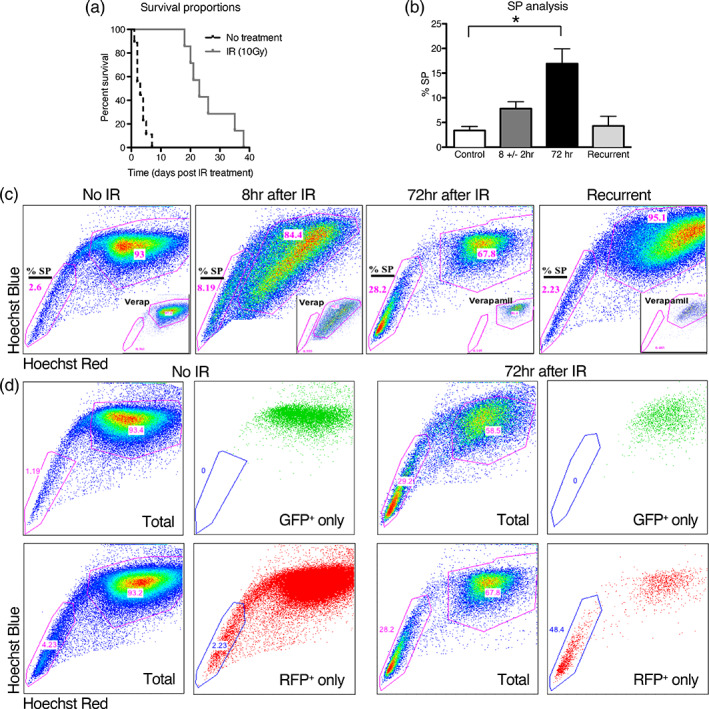
Side population (SP) analysis of a PDGF‐driven mouse model of glioblastoma at early time points after radiation shows that stem‐like cells of the SP are relatively radioresistant and are enriched. (a) A Kaplan–Meier plot showing that mice treated with 10 Gy lived a median of 20 days longer than untreated mice. (b) SP analysis, using Hoechst 33342 dye exclusion assay, of tumors from mice harvested at symptom onset or 8 or 72 hr after IR shows a higher percentage of cells in the SP at 72 hr as compared to control. Error bars represent the SE of the mean. (c) Representative flow cytometry plots, as quantified in (b). SP cells are poorly stained by Hoechst dyes due to efflux pump dye removal, whereas the main population (MP) is highly stained. The percentage of SP cells is highest at 72 hr after IR, but returns to the same level as the control at recurrence. Insets show treatment prior to SP analysis with verapamil as a control, which inhibits the efflux pump, abrogates the SP, and confirms the SP analysis gating strategy. (d) Flow cytometry plots of tumors without and at 72 hr after IR showing SP analysis of Olig2‐expressing tumor cells (GFP+) and tumor cells derived from the earliest tumor cells (RFP+). SP analysis of all tumor cells (Total) are shown prior to gating for GFP positivity (GFP only, top row) or RFP positivity (RFP only, bottom row). GFP+ cells are exclusively in the MP without and at 72 hr after IR, and RFP+ cells are heavily enriched at 72 hr after IR in the SP as compared to control
