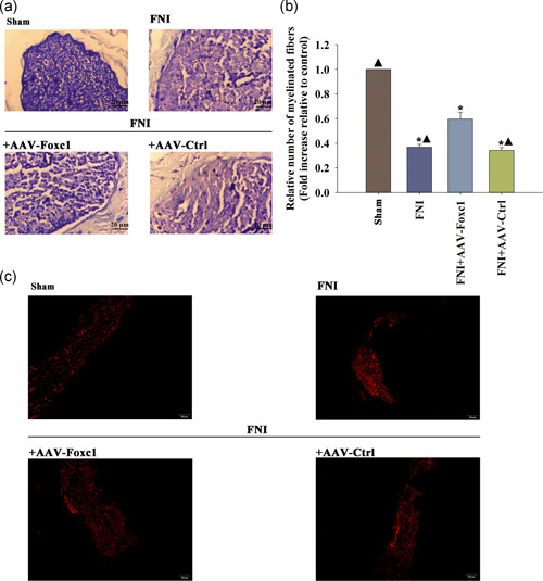
Figure 7.

Effects of transfection with Foxc1 on myelination and Schwann cell (SC) migration in vivo. (a) Analysis of the regenerated myelinated nerve fibers. Cross‐sections from the middle portion of regenerated nerves were stained with toluidine blue at 4 weeks after surgery. (b) The total number of regenerated myelinated nerve fibers. n = 6 per group. Data represent the mean ± SD; Each column represents the mean ± standard deviation (SD) from four independent experiments; *p < .05 versus sham; ▴ p < .05 versus FNI+AAV‐Foxc1. (c) Representative images of the immunofluorescence staining of S100‐β1 in a sagittal section of the FN at 4 weeks after FNI. n = 6 per group. FNI, facial nerve injury
