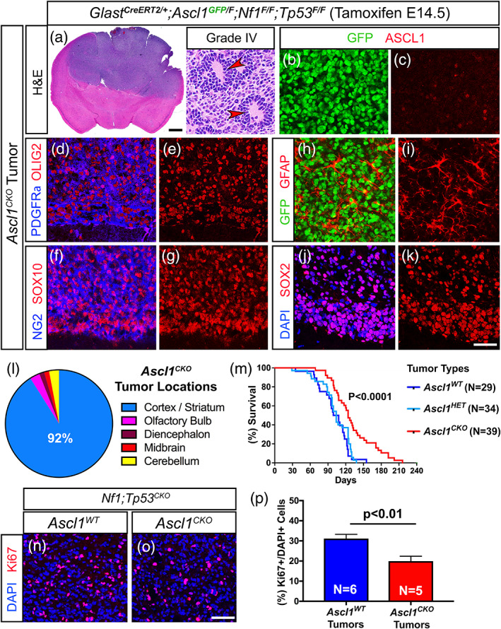
FIGURE 5.

Survival of glioma tumor bearing mice is increased in the absence of ASCL1. (a) H&E staining of an Ascl1 CKO tumor exhibiting pseudopalisading cellular features of Grade IV glioma (arrowheads, insets). (b–k) Immunofluorescence of Ascl1 CKO tumor. GFP, driven by an Ascl1 GFP knock‐in allele, is present in tumor cells but ASCL1 is absent (b and c), indicating efficient deletion of Ascl1 Floxed allele. Expression of OLIG2 and PDGFRα (d and e), SOX10 and NG2 (f and g), GFAP (h and i), and SOX2 (j and k) are unaffected. (l) Incidence of Ascl1 CKO tumors observed in the different brain regions. Over 90% of tumors are found in the cortex and striatum area similar to Ascl1 WT tumors. (m) Survival curve of Ascl1 CKO versus Ascl1 HET tumor mice. Median survival is significantly improved for Ascl1 CKO (122 days) compared to Ascl1 HET (104 days) tumor mice (dotted lines). Note that survival of Ascl1 HET is very similar to Ascl1 WT tumor mice (note blue line is the same as Figure 4p). (n–p) Immunofluorescence (n and o) and quantification of the percentage of Ki67+/DAPI+ tumor cells (p) for Ascl1 WT and Ascl1 CKO tumors. Scale bar is 1 mm for whole brain section and 30 μm for insets in (a); 25 μm for (b–k); and 50 μm for (n and o) [Color figure can be viewed at wileyonlinelibrary.com]
