
Fig. 4.
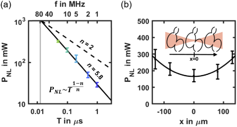
Scaling law of nonlinear photodamage. (a) Nonlinear photodamage threshold PNL in mCherry labeled zebrafish hearts (N = 21 embryos) depending on the laser pulse frequency f = 1/T. Black line shows the result of the scaling law fitted on logarithmic scaled data. The PNL (T) follows a scaling law of order n∼5.8 (see Table S2 in Supplement 1 (1.7MB, pdf) for details). Black dashed line indicates a scaling low of order n = 2 to show how it deviates from 2PEF signal. (b) PNL as a function of the relative position of the heart and illumination beam focus at f = 10 MHz (N = 3 embryos per position). Black line indicates the result of a Gaussian fit. Error bars indicate standard deviation.