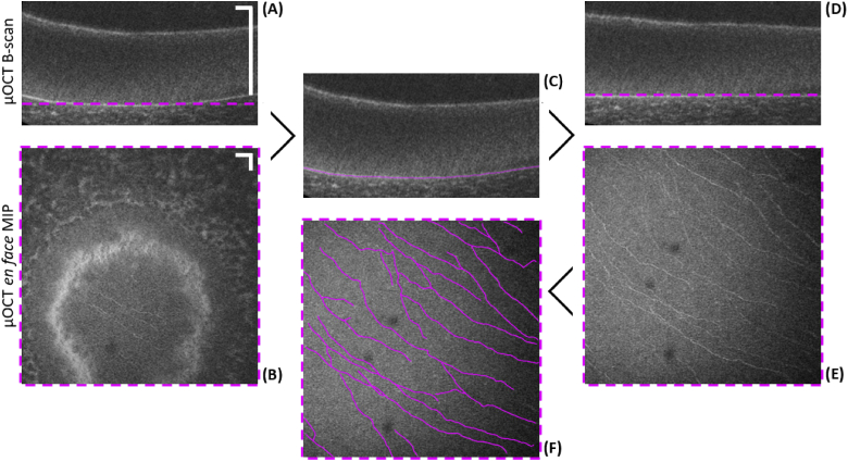
Fig. 2.
Illustration of image segmentation procedure to extract the SBP in a volumetric µOCT scan of a swine cornea. (A) Unsegmented B-scan of corneal epithelium. (B) Unsegmented en face MIP over ∼3 µm in depth at BM. Only a small fraction of the FOV visualizes the SBP. (C) Same B-scan as in (A) with the indicated segmentation line of BM. (D) B-scan segmented and flattened in accordance to the segmentation line. (E) Segmented en face MIP over ∼3 µm in depth just above BM. Entire FOV visualizes the SBP. (F) Results of semiautomatic nerve tracking procedure on the en face MIP of (E). Scale bars: 50 µm.

