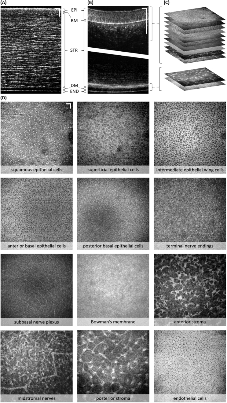
Fig. 3.
Cellular and nerval morphology of epithelial, stromal, and endothelial corneal layers of an excised non-human primate cornea. (A) B-scan of entire corneal thickness (∼400 µm) acquired using a low NA objective lens. Corneal layers indicated: EPI – epithelium, BM – Bowman’s membrane, STR – stroma, DM – Descemet’s membrane, END – endothelium. (B) B-scans of epithelial and endothelial region acquired using high NA objective (stitched together to allow for comparison to (A); vertical scales of (A) and (B) do not match). (C) Illustration of multiple en face MIP of different depth layers computed from the 3D data set. (D) En face MIP over ∼3 µm in depth of twelve specific corneal layers. Scale bars: 50 µm.

