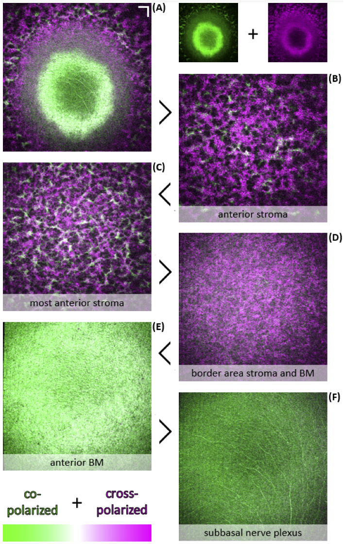
Fig. 6.
Anterior corneal PS-µOCT image data of an excised non-human primate cornea. (A)-(F) Additive color ratio en face MIP of the anterior stroma, BM, and the posterior epithelium (over ∼3 µm in depth). (A) Unsegmented additive color ratio en face MIP of Figs. 5(F) and (G). Anterior stroma (B), most anterior stroma (C), border area between stroma and BM (D), BM (E), and subbasal nerve plexus (SBP) (F) additive color ratio images (co-polarized in green; cross-polarized in magenta; areas of similar intensity result in white). Scale bars: 50 µm.

