. 2020 Oct 23;40(7):1848–1865. doi: 10.1148/rg.2020200159

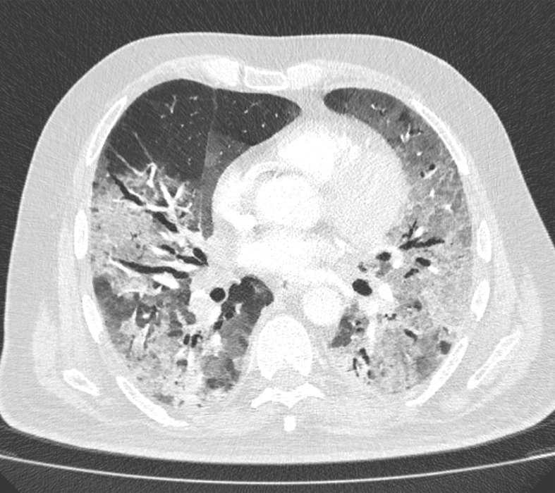
Figure 14b.

PE in a 73-year-old man with COVID-19. (a) Axial nonenhanced CT image (lung window) at baseline shows peripherally diffuse ground-glass opacities in both lungs. (b) Axial contrast-enhanced CT image (lung window) obtained after 10 days shows increased consolidation in both lungs. Note the bronchial dilatation within involved portions of the lungs. (c, d) Axial contrast-enhanced CT image (mediastinal window) (c) and sagittal reconstruction (d) obtained 10 days after the baseline images show a filling defect (arrow) in a segmental pulmonary artery branch in the right lower lobe, consistent with PE.

PE in a 73-year-old man with COVID-19. (a) Axial nonenhanced CT image (lung window) at baseline shows peripherally diffuse ground-glass opacities in both lungs. (b) Axial contrast-enhanced CT image (lung window) obtained after 10 days shows increased consolidation in both lungs. Note the bronchial dilatation within involved portions of the lungs. (c, d) Axial contrast-enhanced CT image (mediastinal window) (c) and sagittal reconstruction (d) obtained 10 days after the baseline images show a filling defect (arrow) in a segmental pulmonary artery branch in the right lower lobe, consistent with PE.