. 2020 Oct 23;40(7):1848–1865. doi: 10.1148/rg.2020200159

Figure 6e.

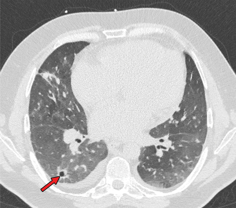
Development of cavitating lung lesions in a 47-year-old man with COVID-19. (a, b) Axial nonenhanced CT images (lung window) obtained at hospital admission show ground-glass opacities in both lungs (early progressive stage). (c, d) Axial nonenhanced CT images (lung window) obtained after 10 days show progressive organizing consolidation (peak stage). (e, f) Axial nonenhanced CT images (lung window) obtained 40 days after the baseline CT images (a, b) show cavitating lesions in both lower lobes (arrow) (late stage).

Development of cavitating lung lesions in a 47-year-old man with COVID-19. (a, b) Axial nonenhanced CT images (lung window) obtained at hospital admission show ground-glass opacities in both lungs (early progressive stage). (c, d) Axial nonenhanced CT images (lung window) obtained after 10 days show progressive organizing consolidation (peak stage). (e, f) Axial nonenhanced CT images (lung window) obtained 40 days after the baseline CT images (a, b) show cavitating lesions in both lower lobes (arrow) (late stage).