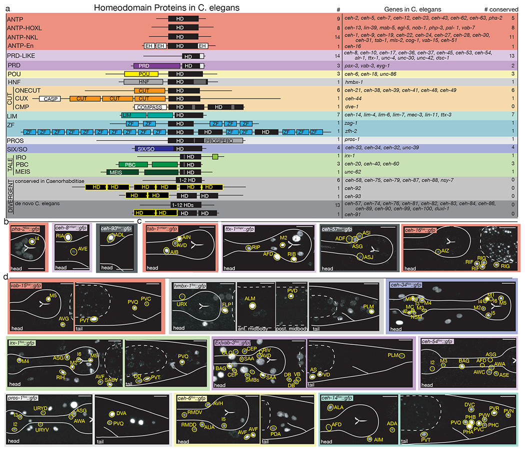
Figure 1: The homeobox gene family in C. elegans and representative expression patterns.

(a) Cartoon representations of homeodomain proteins and their associated domains by subfamily. Numbers of homeobox genes in C. elegans, names of homeobox genes in C. elegans, and number of conserved homeobox genes in humans are based on 2. HD = homeodomain. Yellow “HD” indicates nematode-specific HOCHOB domain, a derivative of the homeodomain 2.
(b-d) Representative images of homeobox genes expressed in 1-2 neuron classes (b), 3-4 neuron classes (c) or 5-18 neuron classes (d). Neurons were identified by overlap with the NeuroPAL landmark strain, outlined and labeled in yellow. Head structures including the pharynx were outlined in white for visualization. Autofluorescence common to gut tissue is outlined with a white dashed line. An n of 10 worms were analyzed for each reporter strain. Scale in bottom or top right of the figure represents 10 um. All other expression patterns are shown in ED Fig.1–8.
