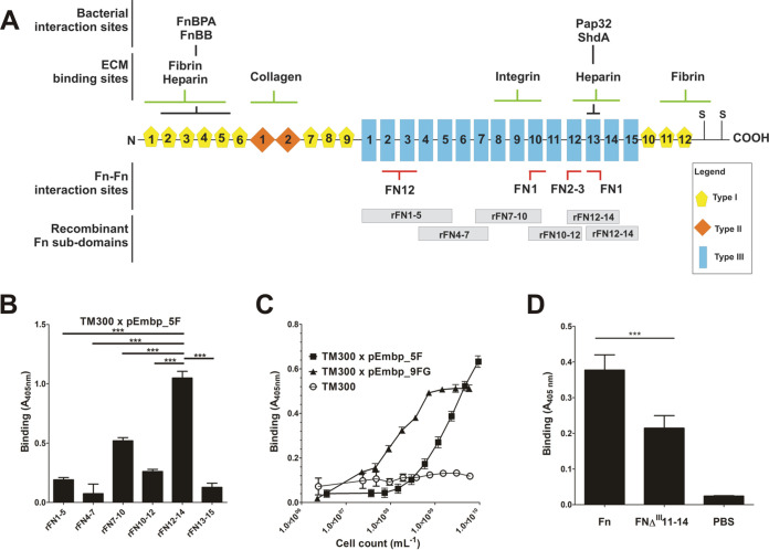
FIG 4.
(A) Schematic representation of the cellular fibronectin monomer. Fibronectin is a 250-kDa multidomain glycoprotein found in various body fluids and various tissues. The type I domain (yellow pentagons) consist of 12 repeats of about 40 aa. Repeats F16 and F17 are intersected by two type II repeats (F21 and F22, each consisting of 60 aa; orange rhomboid), forming the type II domain. In total, at least 15 Fn type III repeats (each consisting of 90 aa) form the type III domain. The secondary structure of type I and type II repeats are stabilized by disulfide bonds. Their absence in type III repeats is related to the elasticity and plasticity of the Fn type III domain. A globular Fn conformation is stabilized by several Fn domain interactions between the two strands of the Fn dimer (e.g., FN12–FN2-3, FN1–FN10, and FN1–FN13; indicated by red bars). Interaction sites with bacterial adhesins (FnBPA [S. aureus], FnBB [S. dysgalactiae], Pap32 [B. henselae], ShdA [S. enterica]), or host extracellular matrix components are indicated by black and green bars, respectively. The positions of recombinant Fn subdomains are indicated by gray boxes. S, position of cysteine residues involved in covalent Fn dimer formation. Extra domains A and B and a variable domain present in plasma Fn are not shown. The figure was adapted from Kubow et al. (46). (B) Adherence of S. carnosus expressing five F-repeats to overlapping Fn type III subdomains. Recombinant Fn type III subdomains rFN7-10, rFN4-7, rFN7-10, rFN10-12, rFN12-14, and rFN13-15 (see panel A) were immobilized on an Immobilizer microtiter plate and incubated with TM300 × pEmbp_5F (108/ml) for 1 h. The unmodified surface served as a control. Adherent bacteria were detected using a polyclonal rabbit anti-S. epidermidis serum and alkaline phosphatase-conjugated anti-rabbit IgG. Bars represent bacterial binding (expressed as the absorption at 405 nm) after background subtraction. Differences between binding to rFN12-14 were significantly different compared to all other recombinant Fn fragments tested (P < 0.0001, Student t test). (C) Adherence of S. carnosus × pEmbp_5F and S. carnosus × pEmbp_9FG to surface-immobilized rFN12-14. A 96-well Immobilizer microtiter plate surface (Nunc, Roskilde, Denmark) was coated with recombinant fibronectin subdomains. Increasing numbers of S. carnosus TM300 × pEmbp_5F and S. carnosus TM300 × pEmbp_9FG were incubated for 1 h on the surface. Adherent bacteria were detected using a polyclonal rabbit anti-S. epidermidis serum and alkaline phosphatase-conjugated anti-rabbit IgG. S. carnosus TM300 wild type served as a control. (D) Adherence of S. epidermidis 1585Pxyl/tetembp to Fn and FnΔIII11-14. Full-length Fn and an isoform lacking type III subdomains FN11 to FN14 (FnΔIII11-14) were purified from supernatants of HEK293 cells transiently transfected with FN-YPet/pHLSec2 or FNΔIII11–14/pHLSec2. Fn isoforms were immobilized on a microtiter plate and incubated with S. epidermidis 1585Pxyl/tetembp grown under embp-inducing conditions. After washing, adherent bacteria were detected using a polyclonal rabbit anti-S. epidermidis serum and alkaline phosphatase conjugated anti-rabbit IgG antibody. ***, significant (P < 0.0001) difference (Student t test).

