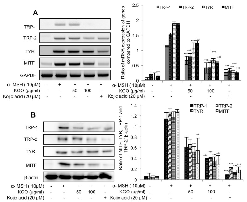
Figure 2.
Suppressed expression of genes in the MITF (microphthalmia associated factor) pathway by KGO in B16/F10 cells. Cells were seeded in 6-well plates, treated with the indicated concentrations of KGO, and then stimulated with α-MSH (10 µM). Total RNA and proteins were extracted, and the TRP-1, TRP-2, TYR, (tyrosinase) and MITF expression levels were assessed by qRT-PCR (A) and Western blotting (B). GAPDH (glyceraldehyde 3-phosphate dehydrogenase) was taken as the internal control for assessing transcriptional expression, and β-actin was taken as the translational control, and all values were compared against them. *** p < 0.001, ** p < 0.05, and * p < 0.01 were considered as statistically significant against the α-MSH-treated group only.

