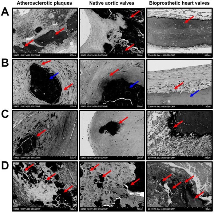
Figure 6.

Mineralisation in the atherosclerotic plaques, calcified native AVs, and failed BHVs. (A) Macrocalcifications within the ECM (indicated by red arrows), magnification 100×. (B) Encapsulated macrocalcifications (capsule indicated by red arrows, calcifications indicated by blue arrows), magnification 500×. (C) Sharp-edged uneven calcifications (indicated by red arrows), magnification 250×. (D) The diversity of calcification patterns (indicated by red arrows), magnification 500×.
