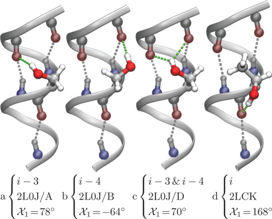
Figure 1.

Different H-bond configurations in solved membrane protein structures, as indicated by the PDB ID and chain (if relevant). Structures a, b, and c are from the M2 H+ channel19 and structure d is from the mitochondrial uncoupling protein 2.20 Note that structures a–c were determined by solid state NMR,19 in which the side-chain conformations were obtained by the refinement procedure. The backbone helical H-bonds are colored in gray, while the bonds formed by the hydroxyl side chain are depicted in green. The χ1 rotamer of the hydroxyl group and the particular H-bond acceptor(s) are noted.
