
Figure 4.
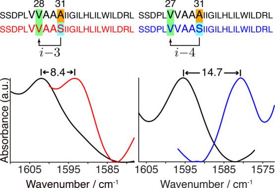
FTIR spectra in the isotope-edited amide I mode region of M2 peptides (Ser22–Leu46, as noted) in hydrated lipid bilayers obtained at room temperature. Amino acids shaded in green are labeled with 13C=18O at position 27 (right panel) or 28 (left panel). The arrows in the sequence depict possible over-coordinated H-bonds by Ser31 (shaded in cyan). Spectra of peptides with an alanine at position 31 (shaded in orange) are depicted in black. The spectra of these peptides with serine at position 31 are depicted in red or blue for peptides labeled at Val28 or Val27, respectively. The spectra were normalized according to each isotope-edited amide I peak.